
Most people consider male and female sexual anatomy to be completely different in every aspect. And there are distinct differences between male and female sexual anatomy, but there are similarities as it relates to sexual structure and function. This lecture we will focus on biological level of analysis. We will examine male and female sexual anatomy and function, along with similarities and differences between the two sexes.
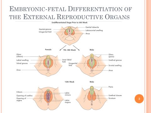
All embryos appear anatomically as female first. If it does not receive certain genetic and hormonal signals, the embryo will continue to develop as female. But when the tissues receive the signal to begin differentiation into a male, the embryonic reproductive organs begin to change their appearance dramatically. Around the 7th or 8th week of gestation, these genetic and hormonal signals will determine the sexual development of the embryo. For female development, you can see that the genital tubercle forms into the glans clitoris, while in male development the genital tubercle forms into the glans penis. The urogenital fold differentiates into the labia minora for females, while in males it develops into part of the shaft of the penis.
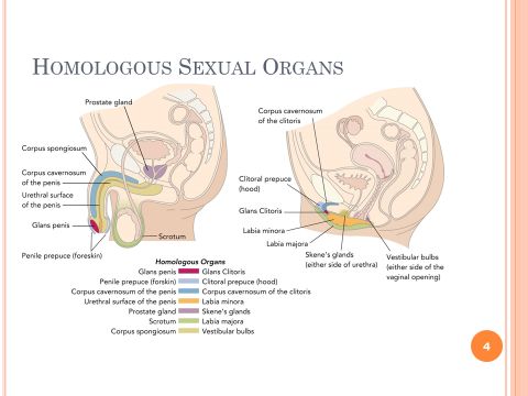
Because the reproductive organs are formed from the same embryonic tissue, males and females have homologous sexual organs. Homologous organs are organs that are similar in position, structure, and evolutionary origin, but not necessarily function. You can see in the developed sexual anatomy between males and females the similarities in the sex organs.
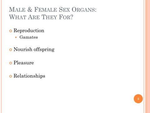
So what's the purpose of male and female sex organs? Clearly the female sex organs serve a reproductive function, but male organs are important for reproduction as well. Male and female sex organs produce and store gametes. Gametes are sex cells of genetic material for reproduction. Male sex organs manufacture the gametes for delivery to the female's reproductive tract. Female reproductive organs have the added ability to provide an environment and nourishment for development of a fetus during gestation and nourishment after birth. The sex organs serve a reproductive function, but they also perform other functions as well. Male and female sexual anatomy brings pleasure, and they also serve to attract potential sex partners. Because of the mutual pleasure partners give each other, it is clear that sexual structures also serve an important role in human relationships.
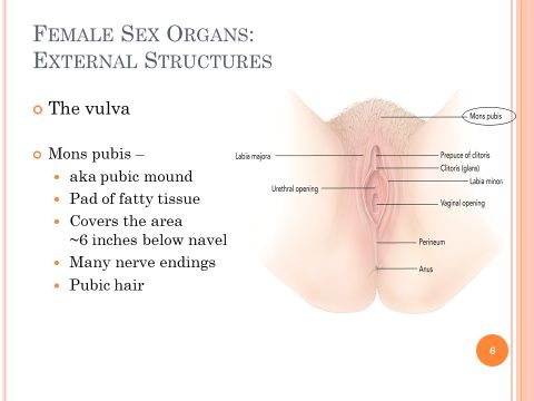
Let's begin by examining the female sexual anatomy. First, we will examine external female sex organs. The external female genitals are the mons pubis, the clitoris, the labia majora, and the labia minora. These structures are collectively known as the vulva. People often use the word vagina when they are actually referring to the vulva. The vagina is an interior structure that we will discuss later. The mons pubis is often referred to as the pubic mound. It is a pad of fatty tissue that covers the area of the pubic bone about six inches below the navel. There is a rich supply of nerve endings in the mons, which can produce pleasure when caressed. From the onset of puberty the mons is covered with pubic hair. The current practice of trimming and shaving pubic hair in America seems mainstream, but it's actually not a new practice. Many cultures for centuries have engaged in this practice for aesthetic or hygienic reasons, such as lack of access to clean water or avoidance of pubic lice. Additionally, many report increased genital sensitivity and partner satisfaction as a reason for pubic hair trimming or shaving now and in the past.
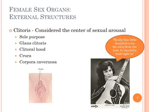
The clitoris is considered the center of sexual arousal. It contains a high concentration of sensory nerve endings. It is very sensitive to stimulation, especially at the tip of its shaft, which is known as the glans clitoris. A fold of skin, known as the clitoral hood, covers the glans when the clitoris is not engorged. Although the glans clitoris is structurally homologous with the glans penis, its sole function is sexual arousal, whereas the glans penis serves the additional function of urine excretion and semen ejaculation. The shaft, or prepuce, of the clitoris is both an external and an internal structure. The external portion is about one inch long and a quarter inch wide. Internally the shaft is divided into two branches called the crura. The crura are the tips of erectile tissue that attach to the pelvic bone and two corpora cavernosa. The corpora cavernosa are hollow chambers that fill with blood and swell during arousal. The hidden erectile tissue of the clitoris plus the surrounding muscle tissue all contribute to the muscle spasm associated with orgasm.
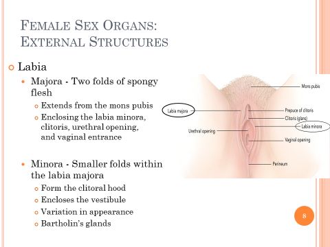
The labia majora, also known as the major lips, are two folds of spongy flesh extending from the mons pubis and enclosing the labia, the clitoris, urethral opening, and vaginal entrance. Within the labia majora are the labia minora, also referred to as the minor lips. These are smaller folds that meet above the clitoris to form the clitoral hood. The labia minora also enclose the urethral and vaginal openings. The areas enclosed by the labia minora is referred to as the vestibule. The labia minora are smooth and hairless, and can vary in appearance quite a bit from woman to woman. During sexual arousal the clitoris becomes erect and the labia minora widen and the vestibule becomes visible. Within the vestibule on either side of the vaginal opening are two small ducts—the Bartholin's glands. These glands secrete a small amount of moisture during sexual arousal.
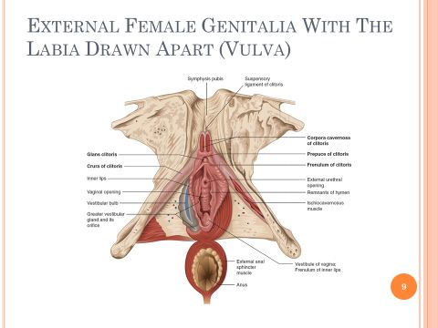
This is an image from your text of the external female anatomy. In this image the labia are drawn apart for a better view of all the structures. Take a minute to go through and identify the structures of the vulva that we just discussed.
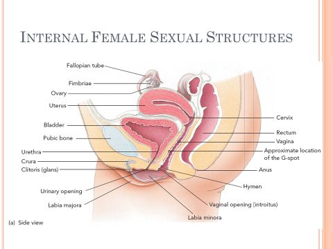
The internal female sexual anatomy and reproductive organs include the vagina, the uterus and its lower opening, the cervix, the ovaries, and the fallopian tubes. We will now discuss each in more detail.
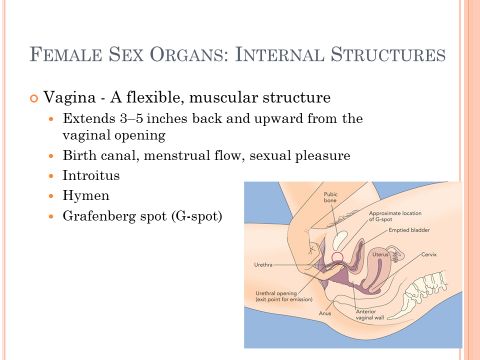
The vagina is a flexible muscular structure that extends three to five inches back and upward from the vaginal opening. It serves as the birth canal through which the infant is born. It also allows menstrual flow to pass from the uterus, and encompasses the penis or other object during sexual expression. When unaroused the walls of the vagina are relaxed and collapse together. However, during sexual arousal the inner two-thirds of the vagina expand, and the pressure from engorgement causes many small blood vessels within the vaginal wall to produce lubrication. Lubrication can occur within 10 to 30 seconds of sexual stimulation. The lower third of the vagina is known as the introitus. This is where the majority of sensory nerve endings are concentrated, and is most sensitive to erotic pleasure and touch. In contrast, the inner two-thirds of the vagina have virtually no nerve endings. This is why a woman cannot feel a tampon when inserted deep into the vagina. Prior to first intercourse or other form of penetration, the introitus is partially covered by a thin membrane containing a relatively large number of blood vessels. This membrane is known as the hymen. The hymen typically has one or several perforations, which allows menstrual blood and mucus secretions to flow out of the vagina. The hymen may be stretched or ruptured by tampon insertion, by the woman's self-manipulation, by a partner during non-coital sexual activity, by accident, or by a healthcare provider conducting a routine pelvic exam. For many cultures, it was or is important for a woman's hymen to be intact on her wedding day as a symbol of purity. The Grafenberg spot, or G-spot, is an area within the vagina reported by many women to be erotically sensitive. The site is located on the front wall of the vagina, midway between the pubic bone and the cervix on the vaginal side of the urethra. The area varies in size from a small bean to half a walnut. It is thought that the close contact between the internal root of the clitoris and the anterior vaginal wall is what leads to the intense and extreme pleasure in women who report sexual enjoyment from contact with the site. However, an exact gland or site has not been found in all women, nor do all women experience pleasure when the site is massaged.
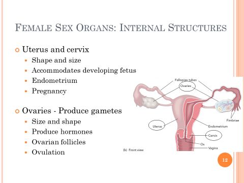
The uterus, or womb, is a hollowed thick wall muscular organ. The tapered end of this pear-shaped organ is known as the cervix. The cervix extends downward and opens into the vagina. If a woman has not given birth, the uterus is about three inches long and three inches wide at the top. For women who have given birth, the uterus is somewhat larger. During pregnancy the uterus expands to the size of a volleyball or larger to accommodate the developing fetus. The inner lining of the uterine wall is known as the endometrium. It is filled with tiny blood vessels. This tissue is built up due to hormonal changes throughout a woman's monthly menstrual cycle in preparation for a fertilized egg. If fertilization has not occurred, the endometrium is shed and expelled through the cervical opening. If fertilization has occurred, the pre-embryo is embedded in the nourishing endometrium. On each side of the uterus is a pair of ovaries. The ovary is a gonad, which is an organ that produces gametes. Female gametes are oocytes. The ovaries are the size and shape of large almonds. In addition to producing oocytes, they serve the important function of producing hormones such as estrogen, progesterone, and testosterone. At birth, the female's ovaries contain about half a million oocytes. During childhood many of these degenerate. Throughout a woman's reproductive years—puberty through menopause—a total of about 400 oocytes mature and are released. The immature oocytes are in a saclike structure called ovarian follicles. At maturation, the follicle ruptures, releasing the oocyte. The release of an oocyte is called ovulation. The egg is viable for fertilization for about 24 hours.
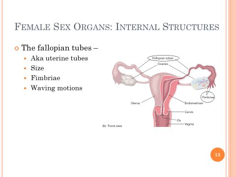
On each side of the uterus are two tubes known as fallopian tubes, or uterine tubes. The tubes are about four inches in length. They extend towards the ovaries but are not attached to them. Instead, finger-like fimbriae drape over the ovary but do not actually touch it. Tiny hair-like cilia on the fimbriae and the ampulla become active during ovulation. Their waving motions and contractions of the walls of the tube transport the oocyte released from the ovary into the fallopian tube.
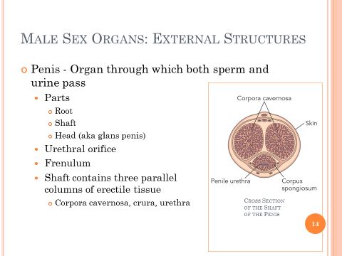
The external male sexual structures are the penis and the scrotum. The penis is the organ through which both sperm and urine pass. It is attached to the male perineum, the diamond-shaped region extending from the base of the scrotum to the anus. The penis consists of three main sections: the root, the shaft, and the head. The root attaches the penis within the pelvic cavity. The body of the penis, or the shaft, hangs free. At the end of the shaft is the head of the penis, which is also referred to as the glans penis. At the tip of the glans is the urethral orifice. The urethral orifice is for both semen ejaculation or urine excretion. On the underside of the penis is a triangular area of sensitive skin called the frenulum, which attaches the glans to the foreskin. The shaft of the penis contains three parallel columns of erectile tissue. The two that extend the front surface are known as the corpora cavernosa. This is the same type of erectile tissue within the crura of the clitoris. The third column, which runs beneath the corpora cavernosa, is called the corpora spongiosum and also forms the glans. Inside the three chambers are a large number of blood vessels through which blood freely circulates when the penis is relaxed. During sexual arousal these vessels fill with blood and expand, which causes the penis to become erect.
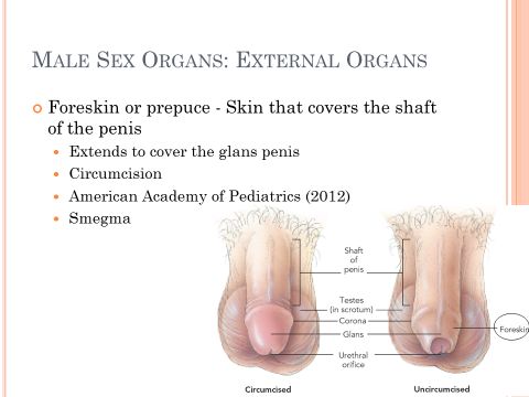
The foreskin, or prepuce, is a loose skin that covers the shaft of the penis and extends to cover the glans penis. The foreskin of a male infant is sometimes surgically removed by a process known as circumcision. As a result of this procedure, the glans penis is left exposed. The reasons for circumcisions are rooted in cultural and religious beliefs more so than any firmly established health principles. There has been much debate in the medical community about the necessity and benefit of this procedure. However, in 2012 the American Academy of Pediatrics, the country's leading pediatric association, released a position statement indicating that the benefits of newborn male circumcision outweigh the risk, and the procedure's benefits justify access to the procedure for families who wish to choose to have it done. Benefits identified were prevention of urinary tract infections, penile cancer, and transmission of some sexually transmitted infections, including HIV. The policy statement is just one factor families should consider when determining to circumcise their newborn son. Beneath the foreskin are several small glands that produce a cheesy substance known as smegma. If smegma accumulates it produces a foul odor that can lead to discomfort and infection. It is important for uncircumcised adult males to observe good hygiene by periodically retracting the skin and washing the glans to remove the smegma.
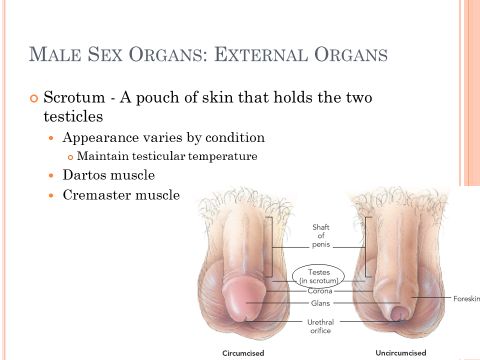
The scrotum is a pouch of skin that holds the two testicles. The skin of the scrotum is more heavily pigmented than the skin elsewhere on the body, and is sparsely covered with hair. The scrotum is divided in the middle by a ridge of skin. The skin of the scrotum varies in appearance under different conditions. For example, when a man is sexually aroused, or when he is cold, the testicles are pulled close to the body. This causes the skin to wrinkle and to become more compact. The changes in the surface of the scrotum help maintain a fairly constant temperature of about 93 degrees Fahrenheit within the testicles. Two sets of muscles control these changes. The dartos muscle is a smooth muscle under the skin that contracts and causes the surface of the skin to wrinkle. The cremaster muscle is within the scrotal sac that causes the testes to elevate.
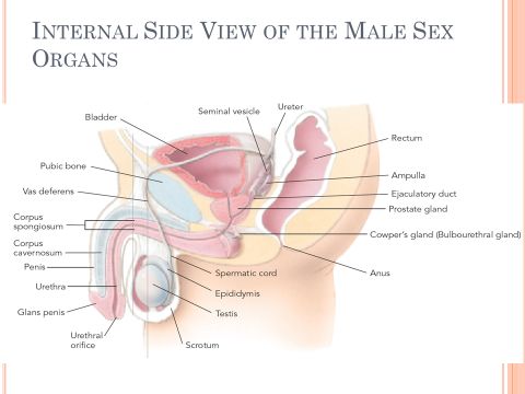
Male internal reproductive organs and structures include the testes, the seminiferous tubules, the epididymis, vas deferens, ejaculatory ducts, seminal vesicles, the prostate gland, and Cowper's glands.
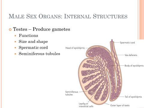
Inside the scrotum are the male reproductive glands, or gonads, which are called the testicles, or testes. The testes have two major functions: sperm production and hormone production. Each testis is about one and a half inches long and one inch in diameter. Each testis weighs about one ounce and is olive shaped. As the male ages, the testes decrease in size and weight. The left testicle generally hangs slightly lower than the right one. Within the scrotal sac each testicle is suspended by a spermatic cord. The spermatic cord contains nerves, blood vessels, and a vas deferens. Within each testicle are about 1,000 seminiferous tubules. These tightly, tiny compressed tubes are about one to three feet long. Within the tubes, spermatogenesis, the production of sperm, takes place.
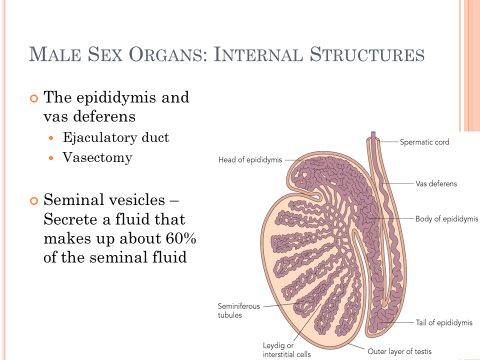
The epididymis and the vas deferens are the ducts that carry sperm from the testicles to the urethra for ejaculation. The seminiferous tubules merge to form the epididymis. The epididymis is a comma-shaped structure consisting of a coiled tube about 20 feet long. In the epididymis is where sperm finally mature. Each epididymis merges into a vas deferens, which is a tube about 18 inches long that extends into the abdominal cavity. The vas deferens joins the ejaculatory duct within the prostate gland. Because the vas deferens can easily be felt within the scrotal sac and is vital for sperm transport, it is usually the point of sterilization for men. The operation is called a vasectomy. A man is still able to ejaculate semen after the procedure; it just no longer contains sperm. At the back of the bladder lie two glands, each about the size and shape of a finger. These are the seminal vesicles, and they secrete a fluid that makes up about 60% of the seminal fluid.
Encircling the urethra just below the bladder is the prostate gland. The prostate gland is the size and shape of a chestnut and produces about 30 to 35% of the seminal fluid in the ejaculated semen. As men age, the prostate enlarges, which can make urination difficult since the prostate encircles the urethra. Other prostate problems range from relatively benign conditions to more serious inflammations and prostate cancer. Below the prostate gland are two pea-sized glands connected to the urethra by tiny ducts. These are the Cowper's or bulbourethral glands. These glands secrete a thick, clear mucus prior to ejaculation, which is often referred to as pre-cum, or pre-ejaculatory. This fluid may appear at the tip of an erect penis. The mucus is alkaline to help buffer the acidity within the urethra and provide a more hospitable environment for sperm. Fluid from the Cowper's glands may contain sperm that have remained in the urethra since a previous ejaculation. Consequently, it is possible for a pregnancy to occur from residual sperm, even if the penis is withdrawn before ejaculation. Therefore the pull out method as a means to prevent pregnancy is not a reliable one.
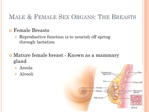
Males and females have some of the same sexual anatomy. Most of these structures do not serve a reproductive function but are often involved in or affected by sexual activities. These structures include the breasts, rectum, and anus. The reproductive function of the female breasts is to nourish offspring through lactation, or milk production. A mature female breast, also known as a mammary gland, is composed of fatty tissue and 15 to 25 lobes that radiate around a central protruding nipple. Around the nipple is a ring of darkened skin called the areola. In response to hormonal signals directly following childbirth, small glands within the lobes called alveoli begin producing milk. The milk passes into the ducts, which open to the outside at the nipple. The amount of milk produced does not vary with breast size because there's very little variation in the amount of glandular tissue among women. In women who are not lactating, breast size depends mainly on fat content, which is determined by heredity.
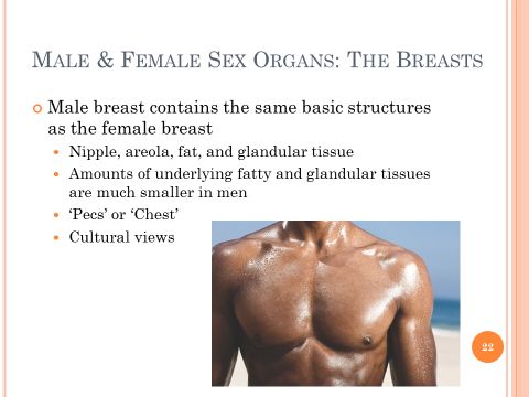
The male breast contains the same basic structures as the female breast—the nipple, areola, fat, and glandular tissue. However, the amounts of underlying fatty and glandular tissues are much smaller in men. But just like women, some men find stimulation of their breasts to be sexually arousing, while others do not. In our culture the male breast is thought of differently than the female breast. Male breasts are often not referred to as breasts, but instead called pecs, or more generally, just the chest. In our society it is acceptable for a man to walk around without a shirt on, but this would be deemed indecent exposure and illegal for women to do so. This is an example of how our culture influences our views on our sexual anatomy.
The anus is the opening of the rectum. It is an organ used primarily for excretion, but can also be used by both men and women during sexual activity. The lining of the rectum is very thin and can tear easily. Because the anus and the rectum do not provide significant amounts of lubrication, most people use some sort of water-based lubricant for penetrative sexual activity. Anal sex involves the insertion of the penis or other object into the rectum. As with vaginal intercourse, anal sex can be potentially unsafe. This is because abrasions of the rectum tissue provide an easy passage for pathogens, such as HIV, to enter the bloodstream. To practice safer sex, partners who engage in anal intercourse should use a latex condom with a water-based lubricant. The anus contains a dense supply of nerve endings that can respond erotically. In men, the prostate gland is located in the front of the rectum, and stimulation of this and nearby structures can be very pleasurable.
In this lecture we examined female and male sexual anatomy and structures. Although female and male genitalia look very different, because they are formed from the same embryonic tissue, they are more similar than often thought.