Table of Contents
- Female & Male Sexual Anatomy Chapters 3 & 4
- Main Topics
- Embryonic-fetal Differentiation of the External Reproductive Organs
- Homologous Sexual Organs
- Male & Female Sex Organs: What Are They For?
- Female Sex Organs: External Structures
- Female Sex Organs: External Structures
- Female Sex Organs: External Structures
- External Female Genitalia With The Labia Drawn Apart (Vulva)
- Internal Female Sexual Structures
- Female Sex Organs: Internal Structures
- Female Sex Organs: Internal Structures
- Female Sex Organs: Internal Structures
- Male Sex Organs: External Structures
- Male Sex Organs: External Organs
- Male Sex Organs: External Organs
- Internal Side View of the Male Sex Organs
- Male Sex Organs: Internal Structures
- Male Sex Organs: Internal Structures
- Male Sex Organs: Internal Structures
- Male & Female Sex Organs: The Breasts
- Male & Female Sex Organs: The Breasts
- Male & Female Sex Organs: Anus
- Summary
Text and Images from Slide
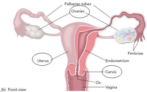
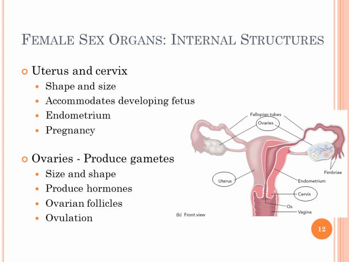
Female Sex Organs: Internal Structures
- Uterus and cervix
- Shape and size
- Accommodates developing fetus
- Endometrium
- Pregnancy<br />
- Ovaries - Produce gametes
- Size and shape
- Produce hormones
- Ovarian follicles
- Ovulation
12